The Dual Excitation with Adaptive Excitation Polygon-scanning Multiphoton Microscope (DEEPscope)

In neuroscience research, imaging neuronal activity in the deep brain is crucial for understanding neurological diseases. However, as imaging depth increases, the field of view usually decreases exponentially. We have improved the fluorescence signal generation efficiency of three-photon microscopy, achieving an imaging field two order of magnitudes larger than traditional three-photon microscopes in deep brain regions with cellular resolution.
Although traditional three-photon microscopes have the capability for deep imaging, their field of view is relatively small. This limitation arises because laser power must be increased with imaging depth, but excessively high laser power can raise the temperature of biological tissue, causing thermal damage. Reducing the field of view lowers the required laser power, thus minimizing the risk of thermal damage.
To overcome the bottleneck of deep, wide field-of-view imaging, DEEPscope incorporates a multi-beam scanning approach and an adaptive excitation module, allowing precise control of laser energy distribution and reducing the laser power needed for imaging. The multi-beam scanning approach splits a single beam into multiple smaller beamlets to monitor neuronal activity, significantly enhancing fluorescence signal generation efficiency, which in turn lowers laser power. The adaptive excitation module focuses laser pulses only on the target imaging area, further reducing required laser power. In traditional multiphoton microscopes, laser energy is usually evenly distributed across the entire sample, including regions not required for imaging. It leads to laser power waste and an increased risk of thermal damage.
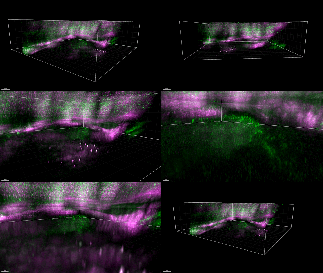
DEEPscope has successfully achieved wide-field neuronal activity monitoring in the deepest cortical layers and hippocampus of mice, expanding the imaging field in the deepest cortical layers by two orders of magnitudes to 3.23 x 3.23 mm. By simultaneously imaging both shallow and deep cortical layers, DEEPscope can record the activity of over 4,500 neurons simultaneously. Additionally, DEEPscope has achieved full-brain imaging in adult zebrafish, capturing structural details over 1 mm deep and more than 3 mm wide, marking the first in zebrafish imaging.
For more details, visit: https://elight.springeropen.com/articles/10.1186/s43593-024-00076-4